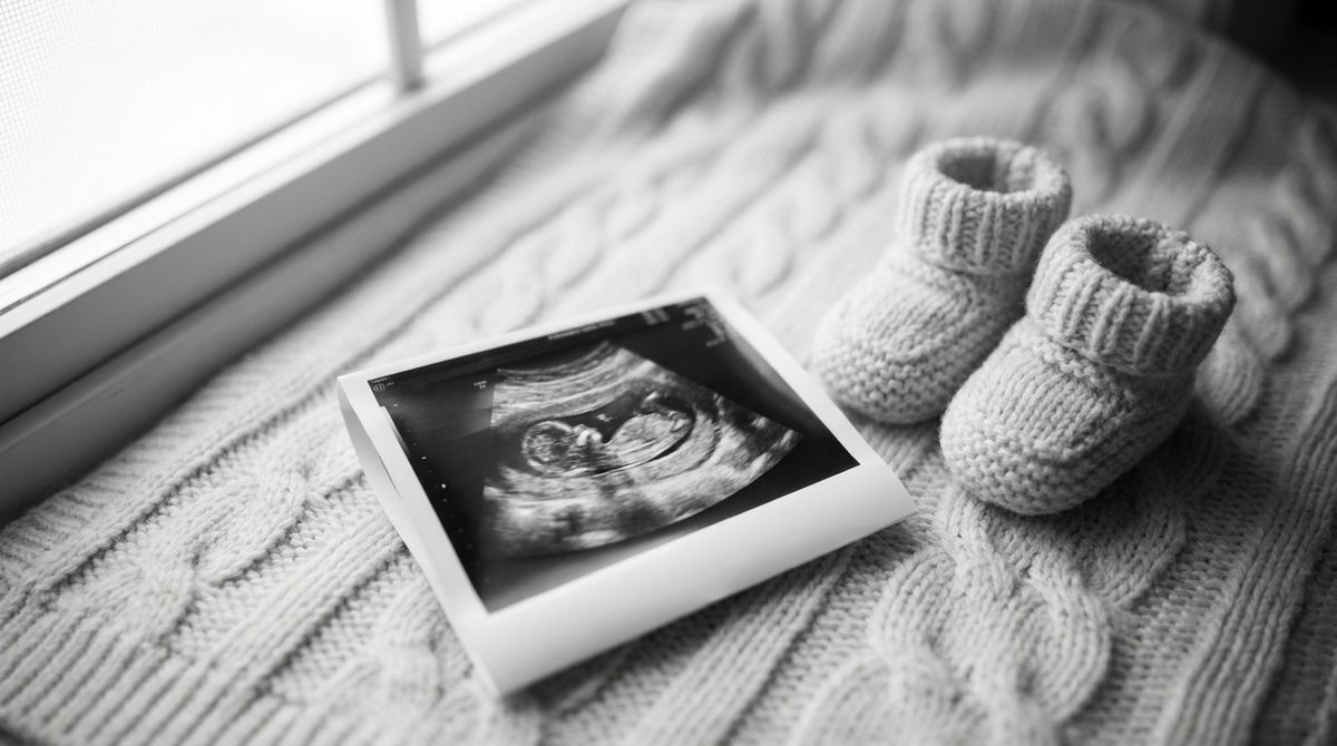
First Ultrasound in Pregnancy: When and What It Shows
Your first pregnancy ultrasound: when it happens, what it shows, the difference between a transvaginal and abdominal scan, and how to prepare.
Mama Ai Team

When the test finally shows two lines, one of the first things you want is to "see" your baby and know that everything is on track. That's exactly what the first ultrasound during pregnancy is for: it confirms the pregnancy, helps establish your gestational age and estimated due date, checks the heartbeat, and shows where the gestational sac has implanted. In this article we'll cover when the first ultrasound is done, what it shows, how a transvaginal scan differs from an abdominal one, how to prepare, and whether it's safe.
First, some reassurance: an ultrasound is a routine, non-invasive part of prenatal care. Most expectant mothers have several scheduled scans during pregnancy, and more often than not they bring good news.
When is the first ultrasound in pregnancy
There's no single "right" day — the timing depends on your situation and the prenatal care protocols where you live. Generally, there are two scenarios.
- An early ultrasound for a specific reason — around 6–8 weeks. This isn't ordered for everyone, only when there's a reason: lower abdominal pain, spotting or bleeding, pregnancy after IVF, an irregular cycle that makes dating difficult, or a suspected ectopic pregnancy. By this point the gestational sac is visible in the uterus, and closer to 6–7 weeks the embryo's heartbeat usually appears — which is why a 6 week, 7 week, or 8 week ultrasound can be so reassuring.
- The first routine (screening) ultrasound — 11–14 weeks. This is part of first-trimester screening, which is recommended for everyone. It's often called the dating scan or, when paired with a nuchal translucency measurement, the NT scan or 12 week scan. It's combined with a blood test to assess the risk of chromosomal conditions.
If your pregnancy is progressing without any worrying symptoms, your first real "meeting" with your baby on screen is often this 11–14 week scan. We explain how gestational age and the due date are calculated in our article How long is pregnancy: weeks, trimesters, and due date. And if you've only just suspected you might be pregnant, you may find our guide to early pregnancy signs before a missed period helpful.

What does the first ultrasound show
Many parents-to-be wonder exactly what the doctor will see on the screen. The first ultrasound during pregnancy answers several important questions at once.
Confirming the pregnancy and its location
The first thing the sonographer checks is whether there's a gestational sac and where it is. Normally it sits inside the uterine cavity. If the gestational sac is located outside the uterus (for example, in a fallopian tube), this is an ectopic pregnancy — a condition that needs urgent medical care. An early ultrasound helps rule it out in time. To learn which symptoms should prompt attention, read about ectopic pregnancy symptoms.
Gestational age and estimated due date
A first trimester ultrasound is the most accurate way to date a pregnancy — which is why this scan is often called a dating scan. The doctor measures the embryo's crown-rump length (CRL) and uses it to calculate your gestational age and due date. The earlier the scan is done (roughly before 13–14 weeks), the more precise the dating — that's why these early measurements are trusted more than the date of your last period, especially with an irregular cycle.
Heartbeat and the embryo's development
A heartbeat can usually be seen from 6–7 weeks, sometimes a little later. If the heartbeat isn't visible yet on a very early scan (say, at 5–6 weeks), that isn't always cause for worry: you may simply be a little earlier along than it seemed. In these cases the doctor will often suggest a repeat ultrasound in 7–10 days to check how things are progressing.
Number of babies
The first ultrasound shows whether one baby is developing or more than one. With a multiple pregnancy, it's important to determine as early as the first trimester whether the babies share a placenta, since that shapes how the pregnancy will be monitored.
Transvaginal vs. abdominal ultrasound — what's the difference
There are two main ways to perform the scan, and which one is used depends mainly on how far along you are.
- Transvaginal ultrasound — a small probe is gently inserted into the vagina. In early pregnancy (6–9 weeks) this gives a clearer picture: the gestational sac, the embryo, and the heartbeat are all easier to see. It's safe and usually painless, though you may feel mild discomfort.
- Transabdominal ultrasound — the probe is moved across your belly after applying gel. This approach is used more often from the 11–14 week scan onward, once the uterus has risen higher.
Sometimes the doctor starts with an abdominal scan and, if needed, switches to a transvaginal one to see the details more clearly. Both methods use the same safe ultrasound waves.
How to prepare for your first ultrasound
No complicated preparation is needed, but a few things will make the scan more comfortable.
- For a transabdominal ultrasound in early pregnancy, you're often asked to come with a moderately full bladder: drink about 0.5 liters (roughly two cups) of water 30–60 minutes before your appointment. A full bladder "lifts" the uterus and improves the view.
- For a transvaginal ultrasound, on the other hand, it's better to empty your bladder right before the scan.
- Wear comfortable clothing that makes it easy to access your belly.
- Bring your maternity notes and the results of any previous scans, if you have them.
Your clinic will give you exact preparation instructions — they may vary slightly.
Is ultrasound safe during pregnancy
This is one of the most common questions expectant mothers ask. An ultrasound uses sound waves, not ionizing radiation (the way an X-ray does). According to major medical organizations, ultrasound is considered safe when it's performed by a trained professional for a medical reason. That's why scans are ordered as often as is genuinely needed for monitoring, and why "keepsake" commercial 3D/4D ultrasound sessions — done purely for souvenir photos without a medical reason — aren't recommended.
The ultrasound schedule during pregnancy
The first ultrasound is only the beginning. Over the course of pregnancy you'll usually have several scheduled screening scans.
- 11–14 weeks — first-trimester screening. This assesses gestational age, nuchal translucency (the NT scan), and early anatomy; the ultrasound is paired with a blood test.
- 18–22 weeks — the anatomy (morphology) scan. This takes a detailed look at your baby's organs, the placenta, and the amount of amniotic fluid. At this stage you can often find out the baby's sex.
- 30–34 weeks — the third-trimester ultrasound. This checks your baby's growth and position and the condition of the placenta, with a Doppler scan (to assess blood flow) added if needed.
Between scheduled scans, your baby's movements become an important "at-home" sign of how things are going. To learn when they begin and what's considered normal, read about fetal movement: when it starts and what's normal.

When to call your doctor
Waiting for your first ultrasound is completely normal in itself. But there are symptoms that mean you should contact your doctor or seek care without waiting for your scheduled appointment:
- heavy vaginal bleeding;
- severe or sharp lower abdominal pain, especially on one side;
- shoulder pain, dizziness, or fainting (these can accompany an ectopic pregnancy);
- a high fever and generally feeling unwell.
These signs don't necessarily mean something is wrong, but it's best not to ignore them and to discuss them with a professional.
Key takeaways
- The first routine ultrasound in pregnancy is usually done at 11–14 weeks (first-trimester screening); an early ultrasound at 6–8 weeks is done only when there's a specific reason.
- It shows whether the pregnancy is present and where it's located, your gestational age and due date, the heartbeat, and the number of babies.
- An early ultrasound helps rule out an ectopic pregnancy.
- In early pregnancy a transvaginal ultrasound is often more informative; later on, a transabdominal scan works well.
- Ultrasound is considered a safe method when it's performed for a medical reason.
This article is for general information only and isn't a substitute for personalized medical advice. For any questions about your pregnancy, please talk to your OB-GYN or midwife.
Sources
Created with AI and reviewed by the Mama Ai team. Educational information — not a substitute for professional medical advice.
We’re with you every week of the way
Download on the App Store